
Bone Marrow: Composition and Hematopoiesis
Heart sounds are brief, transient sounds produced by valve opening and closure and by movement of blood in the heart. They are divided into systolic and diastolic sounds. In most cases, only the first (S1) and second (S2) heart sounds are heard. These are high-frequency sounds and arise from mitral and tricuspid valve closure (S1), as well as aortic and pulmonary valve closure (S2). The third heart sound (S3) may be physiologic (e.g., athletes) or pathologic (e.g., congestive heart failure), and is related to abnormally rapid deceleration of early diastolic left ventricular inflow. The fourth heart sound (S4) is associated with contraction of the atria into partially-filled and non-compliant (stiff) ventricles. S4 is a pathologic sign in the young, but may be found in older individuals due to an age-related decrease in ventricular compliance. Additional sounds include murmurs (physiologic and pathologic), clicks, and snaps. These sounds are heard in individuals with structural abnormalities of the heart such as septal defects, valvular stenosis, and mitral regurgitation.
Overview
Heart sounds
- Heart sounds are produced by:
- Valve opening and closure
- Movement of blood in the heart
- The more turbulent the flow, the more audible the created vibrations.
- On auscultation, 2 heart sounds heard from a normal heart are reflective of the cardiac cycle.
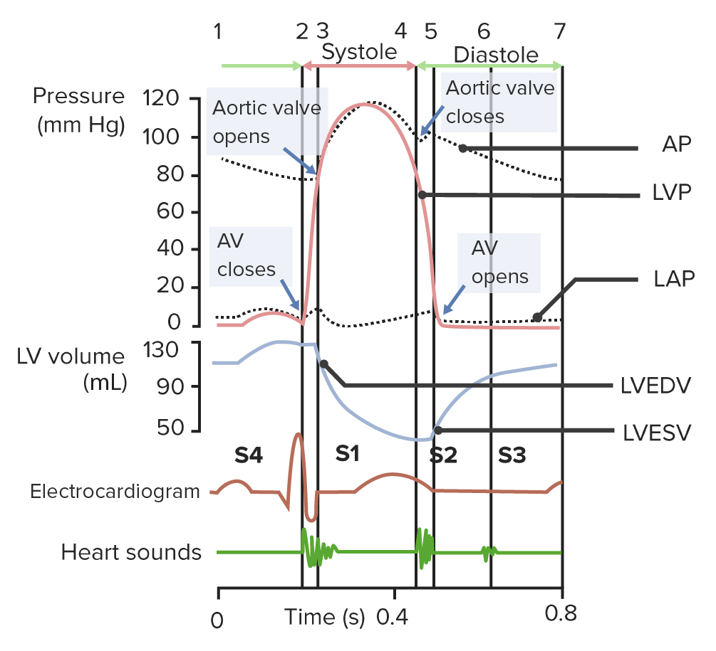
- The cardiac cycle is a sequence of pressure changes in the heart, resulting in:
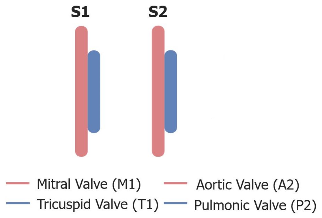
- S1 and S2 mark the beginning and end, respectively, of the cardiac cycle phases: systole and diastole; they are high-frequency sounds.
- Colloquially referred to as the “lub-dub” sound of the heart
- S3 and S4 are low-frequency sounds which may be heard in various conditions.
| Sound | Timing | Association |
|---|---|---|
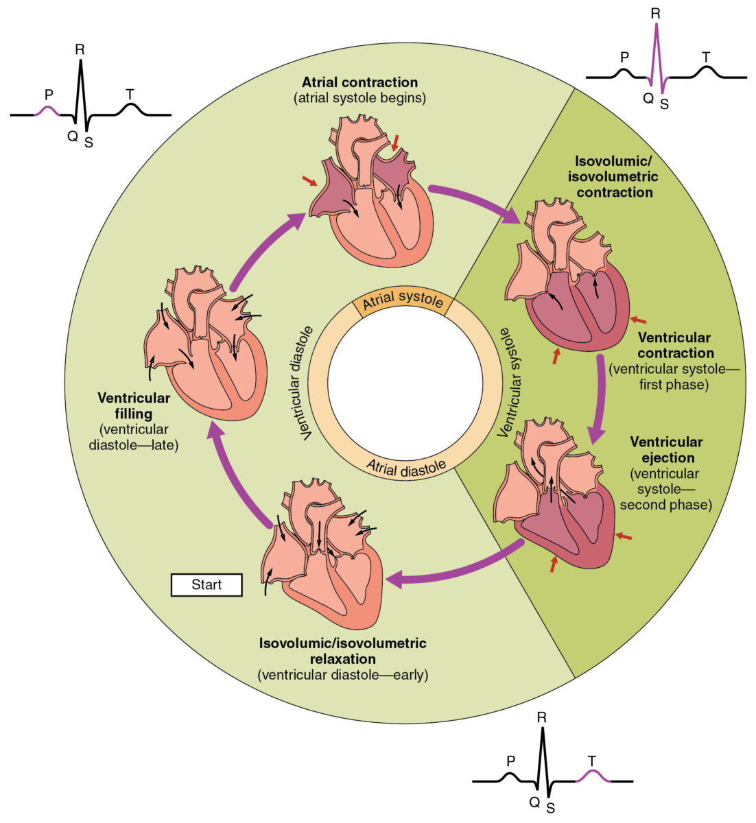
| S1 | Isovolumetric contraction (beginning of systole) | Closure of atrioventricular valves |
| S2 | Isovolumetric relaxation (beginning of diastole) | Closure of semilunar valves |
| S3 | Rapid filling of ventricles (early diastole) | Normal in pregnant women, children, athletesVentricular dilation (e.g., congestive heart failure) |
| S4 | Late filling of ventricles by atrial contraction (late diastole) | Noncompliant or stiff ventriclesPathologic in children and young peopleMay be seen in older people with age-related stiff ventricles |
S1 and S2
S1
- Closure of theatrioventricular (AV) valves
- Mitral valve (M1) closes before the tricuspid valve (T1).
- Indicates the start of systole
- Coincides with the QRS complex and isovolumetric contraction (beginning of systole)
- Loudest over the mitral area (cardiac apex)
S1:
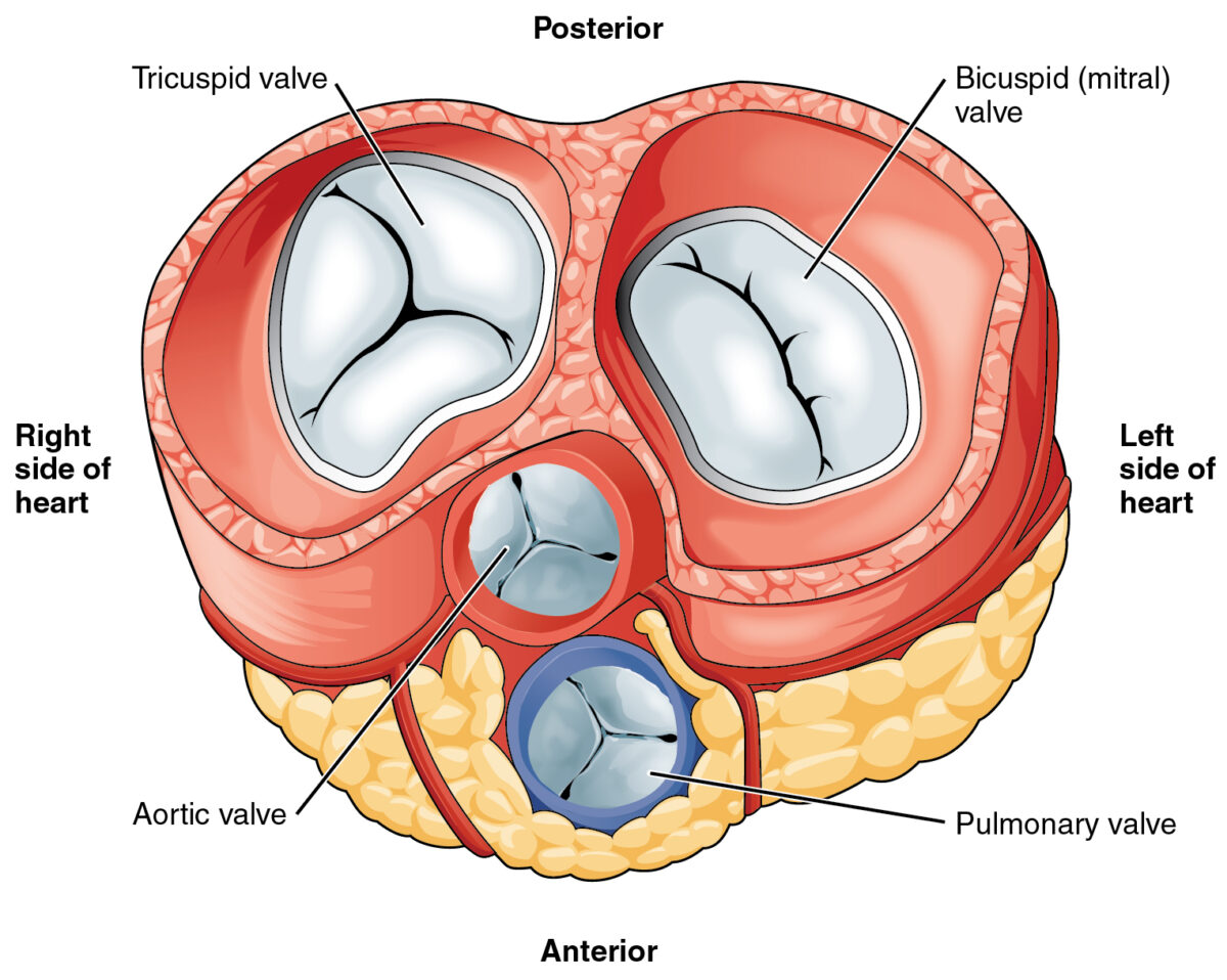
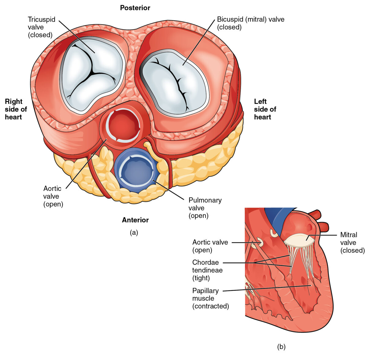
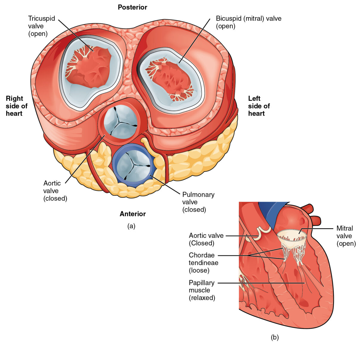
Closure of the atrioventricular valves (tricuspid and mitral) at the beginning of systole. In the systole phase of the cardiac cycle, the right and left ventricles develop pressure, leading to ventricular contraction and ejection of blood into the pulmonary artery and aorta, respectively. Thus, the pulmonary and aortic valves are open. The closed atrioventricular valves prevent the backflow of blood into the atria during ventricular contraction.Image: “2013 Blood Flow Contracted Ventricles” by OpenStax College. License: CC BY 3.0
S2
- Closure of the semilunar valves (pulmonary and aortic)
- Indicates the start of diastole
- Occurs just after the T-wave and coincides with isovolumetric relaxation (beginning of diastole)
- The aortic valve (A2 component of S2) closes before the pulmonic valve (P2 component of S2).
- Loudest at left upper sternal border
Audio:
Normal S1 and S2: In this audio clip, normal S1 and S2 heart sounds can be heard. S1 corresponds to the closure of the AV valves, marking the beginning of
. S2 corresponds to the closure of the semilunar valves, marking the beginning of
.
Report mistake